RESEARCH
My research focus on medical image analysis. Our previous research includes automatic Wireless Capsule Endoscopy (WCE) image diagnosis, brain network analysis and liver lesion detecion. Right now I also focus on prostate cancer classification.
Automatic WCE Image Diagnosis
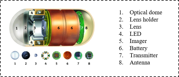
Gastrointestinal (GI) tract diseases, especially in the small intestine and colon, pose the greatest threat to the public health in the world. The GI disease may last for several years to decades, therefore early detection and removal of polyps or ulcers can reduce the chances of disease. WCE has become an irreplaceable tool for diagnostic inspection of the GI tract. It offers a non-invasive alternative to traditional endoscope and enables physicians to explore the GI tract with direct visualization, which is otherwise impossible. We focus on two topics associated with the WCE video clips: abnormality recognition and key frame extraction.
Brain Network Analaysis
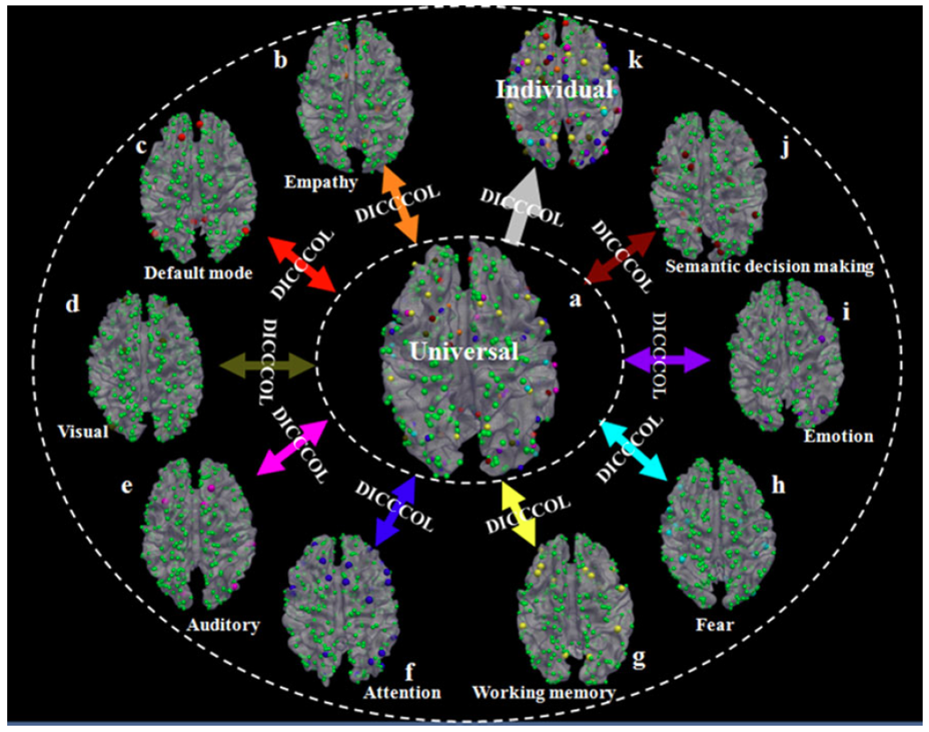
For decades, the human brain mapping community has been interested in defining an anatomically and/or functionally annotated brain atlas and then warping it into individual brains via image registration methods. This atlas warping methodology has dramatically advanced our understanding of the structure and function of the human brain at both individual level and group level. We aim to discover a dense and consistent map of cortical landmarks, named Dense Individualized and Common Connectivity-based Cortical Landmarks (DICCCOLs), each of which is defined by group-wise consistent white-matter fiber connection patterns derived from diffusion tensor imaging (DTI) data. So we aim to take the advantage of existing literature in the BrainMap database to examine the possible functional roles of DICCCOLs via meta-analysis. Moreover, we do analysis about human brain network by investigating the relationship among folding pattern, thickness and fiber density, and assesing graph models for the description of brain networks.
Prostate 3D Image Segmentation [Online Demo]
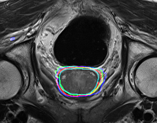
Prostate cancer is the most commonly diagnosed non-cutaneous cancer in men in many parts of the Western world and is a major cause of cancer-related death internationally. Multi-parametric magnetic resonance imaging (mp-MRI) is emerging as a clinically useful tool for detecting and localising prostate cancer. Our target is to develop novel deep learning based models for automatic prostate cancer region segmentation, cancer grading and survival prediction.
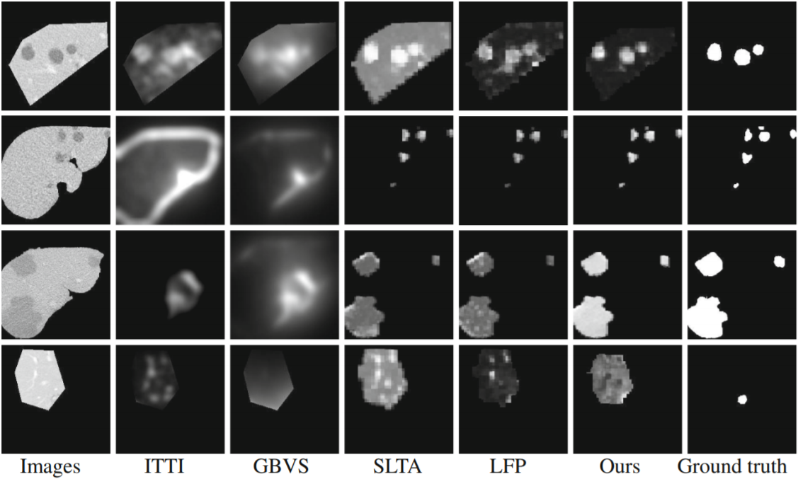
Liver Image Analysis
Liver cancer is a leading cause of cancer deaths worldwide, accounting for more than 600,000 deaths each year. CT is a common non-invasive screening method for detecting liver lesions. However, the large numbers of images in routine liver CT studies, in addition to their high diversity in appearance, have been hurdles for detecting all lesions by visual inspection. Therefore, computer-aided systems play an important role to assist radiologists in automatically detecting liver lesions. Our research tends to segment the live region from CT images, detect the liver lesion and classify liver lesions into three classes: cysts, metastases, hemangiomas.