Medical Applications of Terahertz Imaging
Sponsored by The Shun Hing Institute of Advanced Engineering
Emma MacPherson, KT Chan and YT Zhang - Department of Electronic
Engineering
Anil T. Ahuja - Department of Diagnostic Radiology
and Organ Imaging
W H Cheung - Deptartment of Orthopaedics and Traumatology
Vincent Wallace, collaborator – TeraView Ltd,
Cambridge
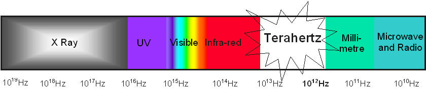
Terahertz (1012 Hz) pulsed imaging is a new technique and has only
emerged in the last five years as a potential new clinical tool for medical
imaging. The optical excitation required for emission of terahertz radiation is
derived from a femtosecond pulsed laser (commonly a
Ti-Sapphire laser at 800nm). The radiation produced is focused onto the sample
of interest and then detected coherently in a reflection
geometry such that the measurement is non-invasive. There are strong water
absorptions in the terahertz region of the electromagnetic spectrum which
therefore means that imaging using terahertz radiation would be a useful tool
to investigate soft tissues.
Terahertz pulsed imaging (TPITM) is a non-destructive, non-ionising
imaging modality with uses in medicine. TPI uses pulses of electromagnetic
radiation typically with a full-width half-maximum of 0.3 ps
and an average power of 100 nW.
The pulses are detected coherently using a photoconductive device and the
Fourier-transformed pulse gives a usable frequency range of 0.1–3 THz. A point
measurement is analogous to an ultrasound A scan.
Reflections off different layers are used to determine the structure at depth
(similar to ultra-sound ‘echoes’). By raster scanning to take several points
within an area (C scan), TPI gives three-dimensional information and can reveal
structures beneath the surface (with spatial resolution precision of 20um and
axial resolution of 40um). A key advantage of terahertz imaging over techniques
such as ultrasound and x-ray is that unique spectral information is obtained
which can be used to distinguish between tissue types. TeraView
Ltd was the first company to develop commercialized terahertz imaging and
spectroscopy systems (www.teraview.com)
and is known worldwide for its research developments in the terahertz field.
Prof. E MacPherson previously worked closely with
Wallace and Fitzgerald at TeraView Ltd, analyzing
breast cancer and skin cancer terahertz spectroscopy data.
There are also many other areas in medicine which would benefit from an
intra-operative probe sensitive to soft tissues. For example osteoarthritis
(OA) is the most common form of arthritis, caused by the breakdown of
cartilage. It usually affects weight-bearing joints like hip, knee, feet and
spine, which causes the joints to degenerate and painful. After cartilage
erosion, bone grinding may occur, leading to thickening and forming of osteophytes. As a result, pain, stiffness, swelling and
reduced range of motion will be presented as symptoms. According to a survey in
2003, the estimated number of people with OA in
In summary, terahertz imaging is a new technique with many potential applications. It uses non ionising radiation to obtain both frequency and time domain information. Therefore it has great potential to be used in medicine to breach the short comings of other medical imaging techniques, and due to the impact of its potential applications, such as reducing the number of second surgical procedures in breast cancer and detecting OA there is a great medical incentive to investigate the in vivo usage of terahertz imaging.